
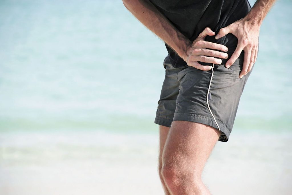
Pain in the lower extremity is an extremely common disorder, which becomes a frequent reason for a visit to a physician. For example, in recent years, the number of visits to a specialist for pain in the knee joint exceeds 4 million (globally). According to some studies, only in the last year, one in three people turned to a specialist with this problem. There are many reasons for this condition: pain in the lower extremities can be caused by both trauma and degenerative changes in the articular surfaces, cartilage abrasion, improper biomechanics of the joints, inflammatory processes, damage to blood vessels and nerves.
Usually, the symptoms of concern indicating the disorder include:
-
Dull, aching pain that aggravates with movement
-
Stiffness or limited mobility in a limb
-
Muscle spasms
-
Difficulty in shifting weight onto one leg
-
Swelling, bruising, or redness of the affected limb.
Before reviewing the causes of such symptoms, let’s dwell on the anatomy of the lower limb and understand which parts of the body can cause pain.

Anatomical features of the lower limb
The lower limb in structure has many similarities with the upper one: it can also be divided into two parts – the belt of the lower limb and the free lower limb. The belt of the lower limb is represented by the pelvic bones and the sacrum, located behind. In fact, the pelvic bones also consist of a combination of three bones: the pubis, ischium and ilium. They are rigidly connected to each other, so they are considered together. The pelvis performs a number of important functions in the human body. First of all, taking into account its shape, the pelvis can be considered as a kind of bowl in which the internal organs lie. This arrangement provides protection of organs from mechanical influences. In addition, one of the functions of the pelvis is due to its participation in the birth process.
The free part of the lower limb is connected to the pelvis through the hip joint: the femur (the upper part of the free lower limb) is attached to the acetabulum formed by the pelvic bones. This joint is cup-shaped and biomechanically resembles a hinge mechanism: during movement of the lower limb, the pelvis remains stationary, while the femur moves in different directions. Movements in the hip joint are normally freely performed due to the sliding of the femoral head along the ilium. Friction is reduced by smooth articular surfaces formed by hyaline cartilage and synovial fluid in the joint cavity, which is known to be produced by the membranes of the joint capsule.
Movements in the hip joint are very extensive, they can be performed in three planes, providing, among other things, a circular motion that is rare for other joints. It is this ability that determines the natural mobility of a person, the ability to walk upright, and the ability to such sports as athletics, gymnastics, etc.
On the other hand, the hip joint, which has such a high functionality, also had weak sides. In particular, due to high mobility, the cartilages of the hip joint are quite fragile, the articular surfaces wear out faster, and inflammation occurs. One of the diseases of the hip joint is coxarthritis. With this disorder, the articular surfaces of the bones that form the joint are deformed, as a result of which the range of motion is dramatically reduced. To prevent this from happening, you need to carefully monitor the health of your joints, exercise wisely, and not overload the musculoskeletal system. A set of useful tips and exercises for the prevention of diseases of the hip joint can be developed by a physiotherapist or a rehabilitation specialist.
Below the hip joint begins the free part of the lower limb. Closest to the place of detachment from the pelvis is the femur. The femur is a long tubular bone that has a number of important anatomical features. The first and most important is the way it connects to the pelvis.

From the upper part of the femur – the greater trochanter, towards the acetabular opening of the pelvis, an outgrowth is found, which plays a connecting role in the system of the lower limb. It has a neck and a head. The femoral neck is a very fragile site, and femoral neck fracture is a common disorder, occurring in 6% of all fractures and affecting 90% of elderly people. The fragility of this part of the bone in elderly is due to the deterioration of blood supply and osteoporosis – a decrease in bone density. Such an injury has serious complications which will be discussed later.
Between the femur and the bones of the lower leg is the knee joint, the largest joint in our body. This joint has a complex structure: three bones and their articular surfaces, a large number of ligaments and synovial bags. Such a complex structure of the joint is due to the fact that it is the junction of the longest levers of the lower limb, which make a large number of movements of a different range in everyday life, in particular, when walking. The knee joint withstands high loads every day, so its shock-absorbing apparatus includes not only the cartilage of the articular surfaces, but also the menisci. Menisci are cartilaginous plates located on both sides of the upper surface of the tibia. They lie between two bones like pillows, softening the excessive load and stabilizing the joint. The integrity of these structures is of great importance. In sports, injuries such as meniscus rupture or injury are common. When this happens, a number of movements, such as going up or down stairs, squats, become almost impossible to perform.
An important part of the knee joint is its ligamentous apparatus. It is represented by lateral (fibular and tibial collateral), posterior (popliteal, arcuate, patellar ligament, medial and lateral supporting) and innerl ligaments. Of the above, four ligaments are responsible for maintaining the stability of the knee joint: anterior cruciate ligament (ACL), posterior cruciate ligament (PCL), medial and lateral collateral ligaments (MCL, LCL). From a biomechanical point of view, the anterior cruciate ligament protects the joint from excessive forward movement, the posterior cruciate ligament protects the joint from backward movement, and the collateral ligaments protect the joint in the frontal plane.
Below the knee joint is the lower leg. The skeleton of the lower leg is represented by two bones – the fibula and the tibia. The second is larger, since it performs a supporting function and withstands the main load. The tibia and fibula are fixedly connected to each other with the help of a flat joint and ligamentous apparatus and act as a whole.
The muscles of the lower leg are divided into anterior, posterior and external groups. The anterior muscle group is responsible for the extension of the foot and toes, adducting the foot. Posterior muscle group, flexes the foot and toes. The outer muscle group abducts and flexes the foot.
Both femurs are involved in the formation of the ankle joint. From the side of the foot, the talus participates in its formation. Movement in this joint is possible around one axis. The structure of the joint and the foot itself ensures an even distribution of the load of the entire body. This joint is quite stable due to the strong ligamentous apparatus, however, its dislocations are a common pathology, which will be discussed below.
Possible causes of pain in the lower extremity
In order to determine the cause of pain in the lower limb, first of all, its localization is specified. Above, we have divided the lower limb into several components, which will now allow us to consider different types of the disorder based on this division.
Pelvic girdle disorder
Speaking about the causes of pain in the lower extremities, it is important to mention such a disorder as the lower crossed syndrome (LCS), when, due to a sedentary lifestyle, a posture is impaired, leading to the muscle imbalance, and as a result, the manifestation of pain, first in the lumbar region, and then in the limbs. This condition significantly affects the quality of life and makes simple daily activities difficult, so it is important to select a treatment with a specialist in time, which will consist of physiotherapy exercises, physiotherapy and manual therapy.

Disorder of the femur and hip joint
Deforming arthritis of the hip joint – coxarthrosis
Coxarthrosis is a degenerative-dystrophic disease of the hip joint, which is caused by damage to the cartilage tissue lining the articular surfaces.
This disease is especially common in traumatology and orthopedics due to the fact that the hip joint can withstand heavy loads, respectively, faster than other joints it undergoes deformation and abrasion of the cartilage lining the articular surfaces. Coxarthrosis can occur for no apparent reason. In this case, it is called primary. In some cases, coxarthrosis occurs as a result of hip dysplasia, trauma, such as a fracture of the femoral neck, dislocations, inflammation and other diseases. Such coxarthrosis is secondary.
Biomechanics and causes of coxarthrosis
The hip joint performs smooth movements in a wide range, and the friction force is reduced due to the hyaline cartilage lining the articular surfaces and the synovial fluid produced by the synovial bag. With coxarthrosis, due to impaired blood supply and dystrophy, the articular surfaces dry out, they become covered with cracks and lose their smoothness. The synovial fluid at the same time thickens, becomes dense. All this increases the friction force in the joint and increases its traumatization, as a result of which, over time, deformation of the bone and cartilage structures develops during movements. In the long term, this staterious consequences, which will be discussed below.
In the long term, this condition causes intermittent or persistent joint pain, limited mobility, and even gait problems (limping).
Risk factors:
-
Constant load on the joint. This load is common in athletes or may be due to excess body weight.
-
In elderly, the blood supply to the joint worsens, which eventually leads to its degeneration and deformation.
-
Circulatory disorders, metabolism, endocrine system.
-
Sedentary lifestyle.
Knowledge of risk factors will prevent the development of the disease, with an individual preventive set of exercises planned by a physiotherapist.
Fracture of the femoral neck
Fracture of the femoral neck is a serious condition, fraught with complications that occurs mainly in the elderly. A fracture of the femoral neck occurs mainly when falling on the side of the house or on the street, and sometimes with minor impacts – an unsuccessful turn in bed, a sharp inclination. Such ease of fracture in the elderly is due to osteoporosis – a decrease in bone density. Clinical picture of the fracture – there is a moderate pain, limited support and mobility of the joint, there may be a mild shortening of the affected limb.
Such fractures can also occur in young people, often as a result of an accident or a fall from a height. An untreated fracture of the femoral neck is often also fraught with nonunion of the bone, and can lead to serious complications, mainly associated with prolonged forced immobility of patients. Over time, bedridden patients may develop congestive pneumonia, bedsores, thrombosis, and depression may develop. It is because of the large number of complications that the mortality rate for hip fracture is 30%.
With a hip fracture, it is important to prescribe the correct treatment. If there are contraindications to surgical intervention, conservative therapy is carried out. Surgical intervention is the restoration of bone integrity with the help of metal structures – osteosynthesis, endoprosthetics.
In the postoperative period and as a preventive measure, a specialist should prescribe a course of special exercises and physiotherapy.
Hip impingement syndrome
Sharp pain in the pelvic region, extending to the thigh, may be associated with infringement of the femoral nerve. In addition to pain, symptoms of infringement include sensory disturbances: paresthesia (creeping sensation) of the front of the thigh, tingling. Muscle twitches may occur.
The cause of the impingement of the femoral nerve is its compression at a certain level. This can occur as a result of muscle spasm, hemorrhage, herniated disc, inguinal hernia, or neoplasm. Most often, the infringement of the femoral nerve occurs in the region of the inguinal ligament (femoral hernia).
Symptoms of damage to the femoral nerve vary from the damage level, and this is due to its anatomical features.
The femoral nerve departs from the lumbar plexus at the level of the 2nd, 3rd and 4th lumbar vertebrae, passes between the major and iliac muscles, descends to the level of the inguinal ligament and exits to the anterior surface of the thigh, where it is divided into branches: skin (sensory), muscular (motor) and saphenous nerve. Accordingly, depending on the level of the damage, the symptoms will differ: they can only capture sensitivity and manifest as paresthesias, they can only affect the motor branches, but most often the infringement occurs at the lumbar level, as a result of which the clinical picture becomes extensive.
Complete neuropathy of the femoral nerve is manifested by paresis of the quadriceps femoris muscle, which is responsible for extension in the knee joint – running and walking are difficult. The leg is fixed in the extensor position. There are pains.
When the femoral nerve is compressed, a consultation with a specialist is necessary to discuss the tactics of treatment, which is mainly conservative, and in some cases may be surgical.
Femoral Anterior Glide Syndrome
Pathogenesis and biomechanics: can develop in people of all ages. It occurs mainly during hip flexion in physical and functional exercise, as well as during running, prolonged squatting, poor posture (for example, flat back), muscle imbalance of the pelvic complex and stabilizing components of the hip joint, as well as hyperextension hips (for example, when running).
Normally, during hip flexion, the femoral head should remain in place or even move slightly backward. With the same syndrome, the head slides forward, causing infringement of nearby soft tissues and nerve endings, while violating the functional and mechanical integrity of the movement. A pain syndrome occurs, which manifests itself not only as a limitation of the range of joint movement, but also in characteristic pains that appear in the anterior region of the thigh, inguinal region, and sometimes radiating pain along the anterior part of the femur.
The reason for this serves as a shortening of the structural elements of the back of the thigh, on the one hand:
Articular capsule, posterior muscle group, posterior pelvic tilt. Along with this, on the other hand, there is overstretching of the anterior capsule, and overstretching of the hip flexor muscle, or its inflammation (Tendinitis), allowing increased anterior glide of the head.
For the treatment and rehabilitation of the syndrome, a special set of exercises has been developed aimed at eliminating its causes.

Disorder of the Knee joint
A common cause of pain in the lower extremity is the knee joint disorder. Above, we discussed its complex structure, from which it can be understood that damage to any of the elements of the joint can cause the disorder and destabilize it.
What are the main causes of the knee joint disorders? Let’s consider the main and most frequent causes.
Knee osteoarthritis
Knee osteoarthritis or gonarthrosis is a chronic degenerative-dystrophic disease characterized by progressive degradation of the cartilaginous apparatus of the knee joint.
As we already know, the cartilage system of the knee joint is normally designed to perform a shock-absorbing and stabilizing function – the knee joint experiences heavy loads, so the body has created conditions for it in which its traumatization is minimal. However, for various reasons, mainly with age, the integrity of the cartilage tissue is impaired, and the depreciation capacity decreases. The knee joint becomes destabilized, friction between its articular surfaces increases, the cartilage structure becomes thinner, which eventually leads to degradation. These changes affect the ability to move freely, joint stiffness occurs, and it becomes difficult to perform daily activities.
Gonarthrosis is the most common form of arthritis. More often, it affects women.
The causes for its development include:
-
Injuries
-
Physical activity
-
Overweight
-
Consequence of previous arthritis
-
Congenital weakness of the ligamentous apparatus а
The disease develops slowly. The first symptoms are mild pain on exertion and a feeling of stiffness. In the later stages, the range of motion in the joint gradually decreases, stiffness develops, pain becomes more intense and appears even at rest. The gait becomes unsteady.
It is important to prevent the progression of gonarthrosis and to choose the right treatment with an orthopedic traumatologist in time, which includes a complex of physiotherapy, manual therapy, and massage.
Injury of the knee menisci
Injury of the knee menisci – an impairment of the integrity of the meniscus located in the cavity of the knee joint. This condition is most often the result of a sports injury during rotation of the lower leg, when the foot remains fixed in place (skating, wrestling, rugby, football). Sometimes the injury is accompanied by a typical “click”. Sometimes such an injury develops with a direct blow to the knee joint (falling onto steps) or a fall from a height onto straightened legs. Injury or rupture of the meniscus rarely goes unnoticed. The acute period begins immediately after the injury. The patient clearly remembers the moment of injury, experiences pain, tries to avoid movements in the joint. The lower leg is fixed in the flexion position. When you try to move, there is a feeling of jamming of the joint – blocking the joint. This condition can become chronic and manifest as periodic pains.
In the acute period, you should refer to a medical specialist for care which consists in eliminating the blockade. During rehabilitation – a complex of exercise therapy and physiotherapy.
Anterior cruciate ligament (ACL) injury. The unlucky triad
The ligamentous apparatus is very developed in the knee joint. Its stability is mainly maintained by four ligaments, one of which is the anterior cruciate. It prevents excessive movement of the lower leg forward and occurs, respectively, in cases where the load on it is too high. Such an injury is common among skiers when the skis get stuck in the snow and the person, fixed in the boots, falls forward. The injury can also occur when falling forward in high boots, hyperextension of the knee, or with a direct blow.
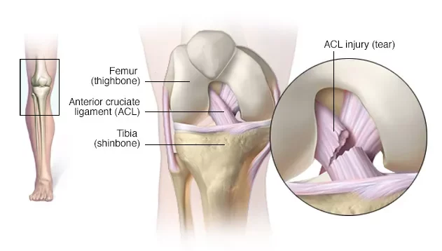
A rupture or injury of the cruciate ligament is accompanied by severe pain and clicking at the time of injury. The patient cannot lean on the leg, the knee swells strongly, symptoms of a “pseudo-block” appear – the impossibility of flexion and extension in the knee joint.
In the acute period of the disease, medical specialists eliminate edema and reduce inflammation. In the recovery period, fixators are indicated – bandages, which allow you to restore joint mobility more effectively with exercise therapy and physiotherapy. The initial set of exercises is indicated in the first hours after the injury, this is important so that joint contracture does not develop. Numerous studies in recent years have proven the effectiveness of conservative treatment -massive rehabilitation physiotherapy, compared with surgery to reconstruct the damaged ligament. Comparing the results of the two interventions in a yearly perspective, the same results were found! Unfortunately, the terms of recovery and return to normal activities are very long (more than 6 months) in both cases.
Unlucky triad is a condition that occurs when the anterior cruciate ligament (ACL), medial collateral ligament (MCL), and meniscus are torn at the same time. The unlucky triad occurs with a side impact to the knee and is common in sports such as football, rugby, hockey, or motocross. In the process of such an injury, the leg, as it were, turns laterally and overextends, which leads to a simultaneous rupture of the above elements.
The simultaneous damage of the menisci + two of the four main stabilizing elements of the knee leads to its destabilization, visible to the naked eye when twisting. Movement is severely limited. In addition to severe pain, there is swelling and stiffness. This condition requires qualified medical attention. A patient needs surgical treatment and restoration of the ligamentous apparatus. An important element of rehabilitation is physiotherapy. Proper treatment will help restore mobility in the joint.
Patello-Femoral Syndrome or chondromalacia patella
The most common cause of pain in the front part of the knee, along with the tendon of the same name.
Chondromalacia is a softening and wear of the cartilage of the lower surface of the patella (the cartilage of the patella is the most vulnerable cartilage) There is a higher incidence in young people than in adults. It is mainly caused by injury, wear and tear caused by excessive stress on the knees, weakness or imbalance of the thigh muscles, as a result which causes the patella to move along an incorrect path, causing damage and abrasion of the cartilage, the appearance of roughness and cracks on its surface.
Instead of a smooth movement between the patella and the lower part of the femur, the patella rubs against the thigh, and the roughness of the cartilage causes pain. Thus, there is an inflammatory process and functional disorders, restrictions on the dynamics of movement.
An important element of rehabilitation is physiotherapy. Adequate treatment and a properly designed set of exercises will help restore mobility in the joint.

Disorder of the ankle joint
Dislocation of the ankle joint. Ankle instability
Ankle dislocation is the most common cause of ankle disorder. It manifests itself in the displacement of the articular surfaces of the tibia, talus and fibula relative to each other. Usually, this condition is caused by a twisted leg or injury, sports activity, an accident. With such injuries, damage to the ligamentous apparatus of the ankle occurs – the calcaneofibular, posterior and anterior talofibular (tucking inward) or deltoid ligament (tucking outward). A patient complains of severe pain after the injury. The joint tissues are edematous, cyanotic, with subluxations moderate deformation is determined. Movement is severely limited. A complete dislocation is a more serious condition, the deformity in this case is significant, often accompanied by a fracture of the ankles. Movement is not possible..
Classification of the degree of damage to the ligamentous apparatus: from stages 1-4, when at the initial stage microtearing of the ligament is found, and in stage 4 we are talking about its complete rupture. Depending on the degree of damage to the ankle ligaments, instability can develop, which over time, without proper treatment and recovery, can lead to repeated dislocations and subluxations – this is the main component that must be taken into account when building a rehabilitation program for the joint and the entire lower limb. It is also important to mention the presence of proprioceptors (specific receptors) responsible for receiving information about the location of the body in space and balance. They are found in large numbers in the ankle joint, and in smaller numbers in other joints of the lower limb. Improving proprioception is an equally important component in building a rehabilitation program for the ankle joint and the entire lower limb.
Instability of the ankle joint develops after dislocations, due to the weakness of the ligamentous apparatus, which almost does not recover after a severe dislocation. The ligament remains in a stretched state and performs its functions of static stabilization worse. Instability can become chronic, with the result that tucking and displacement of the structures of the foot occur regularly. Chronic instability of the ankle joint develops gradually.
There are 3 stages of instability:
-
Function of the ankle joint is preserved, the pain is mild.
-
Function is broken. Severe swelling of the joint, stiffness of movements, pain during exercise and at rest.
-
Ligaments are completely torn. Severe pain when trying to move, swelling.
For the treatment and rehabilitation of dislocation of the ankle joint, as well as the development of preventive measures to prevent chronic instability, it is required to strengthen the muscular, compensatory-stabilizing apparatus, which will perform dynamic stabilization of the joint. Therefore, a detailed, professional development of specific sets of exercises by a specialist physiotherapist and a rehabilitation doctor is necessary.
Ankle fracture
Ankle fracture usually accompanies a complete dislocation of the ankle and is the result of a strong twisting of the leg. Middle-aged and elderly people are especially susceptible to such injuries – this is due to the peculiarities of coordination of movements and physical form. Ankle fractures are especially common in the cold season, when ice appears. In some cases, it may be due to compression of the ankle as a result of an accident or a heavy object falling on the leg.
The fracture is accompanied by pain, swelling, limitation of support and movement. The severity of symptoms depends on the degree of damage to the ligamentous apparatus, there is a clinic of dislocation of the ankle. There is also a “symptom of irradiation” – pain in the ankles during compression of the bones of the lower leg in the middle third.
Reposition of fragments and plaster immobilization are indicated as treatment. Physiotherapy is recommended during the recovery period.
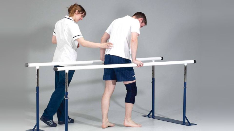
Video: “Exercises for the Lower Limb Problems“
Exercises for the pelvic girdle and hip joint. Set No.2 is aimed at strengthening the muscles
For additional information about the sets of exercises for rehabilitation of the pelvic girdle and hip joint, knee joint and ankle joint, you can watch a video demonstrating exercises and rehabilitation recommendations.
Treatment of the lower limb disorder
The purpose of a particular treatment method always depends on the cause of the pain. In any case, it is necessary to consult a specialist who will establish the cause of the pain through diagnostic tests (MRI, X-ray), physical examination and tests, clarifying the complete clinical picture. After the diagnosis is established, the treatment of the problem begins. In most cases, methods of rehabilitation and prevention of the progression of diseases of the lower limb are effective. A rehabilitation and gradual recovery program can be selected by a physiotherapist or rehabilitation specialist. Therapeutic exercise is an integral part of treatment to restore limb mobility, as well as physiotherapy or manual therapy.

Check out the demo version of our sets of exercises for the Lower Limb Problems on YouTube
Our website presents the following sets of exercises for rehabilitation of the pelvic girdle and hip joint, knee joint and ankle joint:
-
EXERCISES FOR THE PELVIC GIRDLE AND HIP JOINT. SET №1 IS AIMED AT MUSCLE STRETCHING (IMPROVEMENT OF FLEXIBILITY OF THE MUSCULAR APPARATUS) AND INCREASE OF THE RANGE OF JOINT MOVEMENT
-
EXERCISES FOR THE PELVIC GIRDLE AND HIP JOINT. SET №2 IS AIMED AT STRENGTHENING THE MUSCLES
-
EXERCISES FOR THE KNEE JOINT. SET №3 IS AIMED AT STRETCHING THE MUSCLES (IMPROVING THE FLEXIBILITY OF THE MUSCULAR APPARATUS) AND INCREASING THE RANGE OF JOINT MOVEMENT
-
EXERCISES FOR THE KNEE JOINT. SET 4A IS AIMED AT STRENGTHENING THE MUSCLES. INITIAL STAGE
-
EXERCISES FOR THE KNEE JOINT. SET 4B IS AIMED AT STRENGTHENING THE MUSCLES. MID STAGE
-
EXERCISES FOR THE KNEE JOINT. SET 4C IS AIMED AT STRENGTHENING THE MUSCLES. ADVANCED STAGE
-
EXERCISES FOR THE ANKLE JOINT. SET №5 IS AIMED AT STRETCHING THE MUSCLES (IMPROVEMENT OF FLEXIBILITY OF THE MUSCULAR APPARATUS) AND INCREASE OF THE RANGE OF JOINT MOVEMENT
-
EXERCISES FOR THE ANKLE JOINT. SET №6D IS AIMED AT STRENGTHENING OF THE MUSCLES AND IMPROVEMENT OF PROPRIOCEPTION AND STABILITY OF THE ANKLE AND ENTIRE LOWER LIMB. INITIAL STAGE
-
EXERCISES FOR THE ANKLE JOINT. SET №6E IS AIMED AT STRENGTHENING OF THE MUSCLES AND IMPROVEMENT OF PROPRIOCEPTION AND STABILITY OF THE ANKLE AND ENTIRE LOWER LIMB. MID STAGE
-
EXERCISES FOR THE ANKLE JOINT. SET №6F IS AIMED AT STRENGTHENING OF THE MUSCLES AND IMPROVEMENT OF PROPRIOCEPTION AND STABILITY OF THE ANKLE AND ENTIRE LOWER LIMB. ADVANCED STAGE
In addition to the listed sets of exercises please check out article and sets of exercises for the Post-traumatic Rehabilitation of Gait Disorders


